A better understanding of the physiological structures that bacterial meningitis affects can allow an individual to understand how and why one would get ill. In simple terminology, Bacterial Meningitis can be described as the inflammation of the meninges, a protective membrane that covers the human brain and spinal cord.
What is the Meninges?
The meninges compose of 3 envelope membranes called the dura mater, arachnoid mater, and pia mater which serve to protect the central nervous system (CNS). The cranial meninges surrounds the brain and the spinal meninges envelopes the spine (Illustrated in Figure 1)[15]
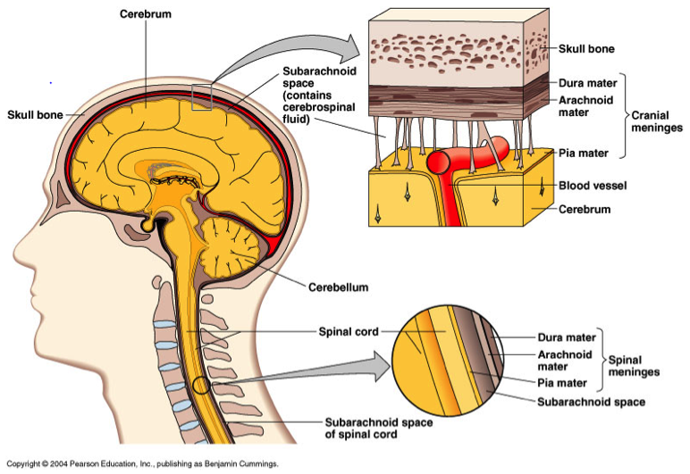
FIGURE 1:
Locations and structures of cranial and spinal
meninges [5]
Pia Mater
The pia mater, a memberane composed of fibrous tissue, is the innermost layer and adheres to the brain or spinal surface. Its exterior side is layered with a sheet of flat cells, which are impermeable to fluid. Blood vessels penetrate through this membrane in order to supply the brain and spinal cord.[15]
Arachnoid Mater
The arachnoid mater is a thin, transparent membrane made up of fibrous tissue and covered by flat cells that are impermeable to fluids. This membrane maintains a loose fitting. Arachnoid trabeculae serve to loosely connect the arachnoid and pia membranes by penetrating the arachnoid membrane and through the subarachnoid space and connecting to the pia matter. Moreover, the subarachnoid space serves as the secretory region in which cerebral spinal fluid flows through. [15]
Dura Mater
This outermost layer of the meninges serves to support the dural sinuses, a venous network which carry blood form the brain to the heat, and is described as a thick, dense, and strong membrane that is made up of dense fibrous tissue. Just like the other two membrane layers, the dura is also covered by flat cells to that are impermeable to fluids. In addition, finger like projections from the arachnoid mater into the dura mater, called arachnoid granulations, allow for the reabsorption of cerebral spinal fluid (CSF) from the sub arachnoid space into the dural venous sinuses (Illustrated in Figure 2).[15]
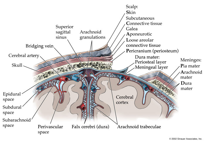
FIGURE 2: Illustrates the arachnoid granulations embedded in the meninges [6]
How cerebral spinal fluid (CSF) circulation works?
CSF has multiple roles which consists of cushioning the brain like a shock absorber, circulates chemical and chemicals obtained from the blood, and removes waste form the brain. It composition consists of 15 - 60 mg/100 mL of protein, 50 - 80 mg/100 mL of glucose, and 0-5 white blood cells. [16]
CSF circulates among the brain ventricles, the spine’s central canal, and the subarachnoid space between the pia mater and arachnoid mater. The production of CSF begins in the choroid plexus which is comprised of ependymal cells, blood vessels (containing endothelial cells), and connective tissue. A tight junction forms between the endothelial and the ependymal cells, thus creating a portion of the blood-brain barrier known as the blood CSF barrier which control what molecules ultimately enter the brain. [19]
The path followed by CSF begins in the lateral ventricle and flows to the third ventricle via the the interventricular foramina. From here the CSF travels through the cerebral aqueduct into the fourth ventricle. Afterwards, the CSF enter the subarachnoid region that surrounds the brain and spinal cord via the lateral apertures or the median apertures. In addition, some CSF enters the spine’s central canal. Reabsorption of CSF is performed by the arachnoid granulations which then allows for the CSF to enter the venous portion of the bloodstream. The rate of this of this process occurs at the rate of about .4 mL/min.(Illustrated in Figure 3)[19]
FIGURE 3: Illustrates CSF circulation [7]
